Home

cervo gooey elegante collagen tem liscio rivista Recuperare

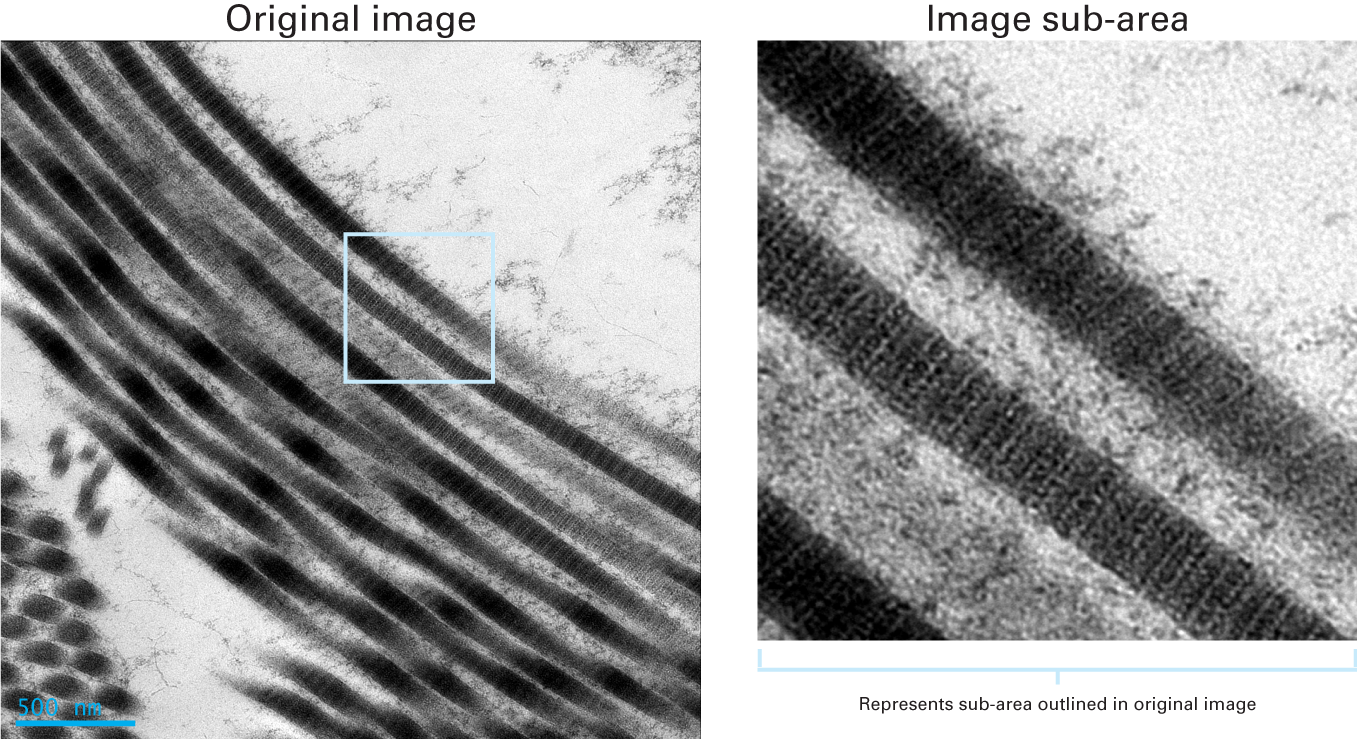
Using transmission electron microscopy and 3View to determine collagen  fibril size and three-dimensional organization | Nature Protocols
Using transmission electron microscopy and 3View to determine collagen fibril size and three-dimensional organization | Nature Protocols

Porcine cornea stromal collagen architechture, TEM, Transmission electron  microscopy | Animal print rug, Printed rugs, Animal print
Porcine cornea stromal collagen architechture, TEM, Transmission electron microscopy | Animal print rug, Printed rugs, Animal print

Collagen Fibres, Tem Photograph by Thomas Deerinck, Ncmir | Pixels

Representative transmission electron microscopic (TEM) images of... |  Download Scientific Diagram
Representative transmission electron microscopic (TEM) images of... | Download Scientific Diagram

Collagen Fibril Diameter by TEM.

Collagen damage - TEM | Wellcome Collection

Clearly resolved collagen fibrils | Gatan, Inc.

Transmission electron micrograph (TEM) showing the typical corneal stroma  or substantia propria. It is composed of layers of collagen fibres, forming  Stock Photo - Alamy
Transmission electron micrograph (TEM) showing the typical corneal stroma or substantia propria. It is composed of layers of collagen fibres, forming Stock Photo - Alamy
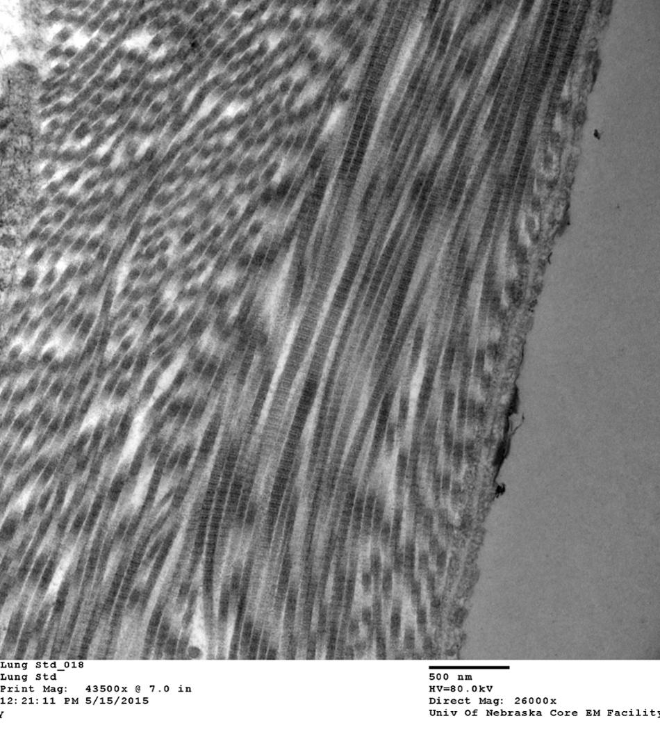
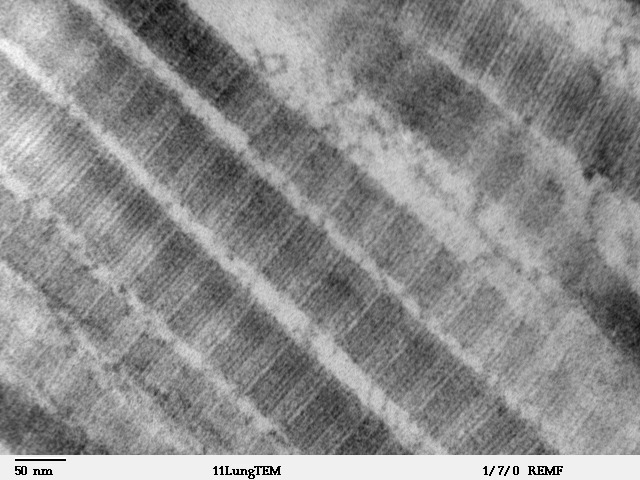
Collagen Fibrils in Lung Tissue | VCR | University of Nebraska Medical  Center
Collagen Fibrils in Lung Tissue | VCR | University of Nebraska Medical Center

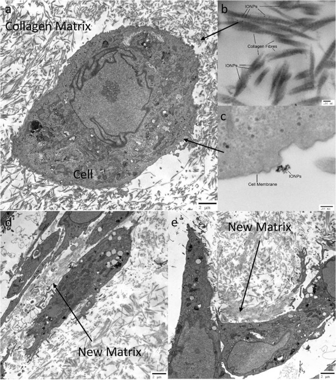
Development of a 3D Collagen Model for the In Vitro Evaluation of  Magnetic-assisted Osteogenesis | Scientific Reports
Development of a 3D Collagen Model for the In Vitro Evaluation of Magnetic-assisted Osteogenesis | Scientific Reports

TEM micrographs showing ultrastructure of collagen fibers assembled in... |  Download Scientific Diagram
TEM micrographs showing ultrastructure of collagen fibers assembled in... | Download Scientific Diagram

Human Structure Virtual Microscopy

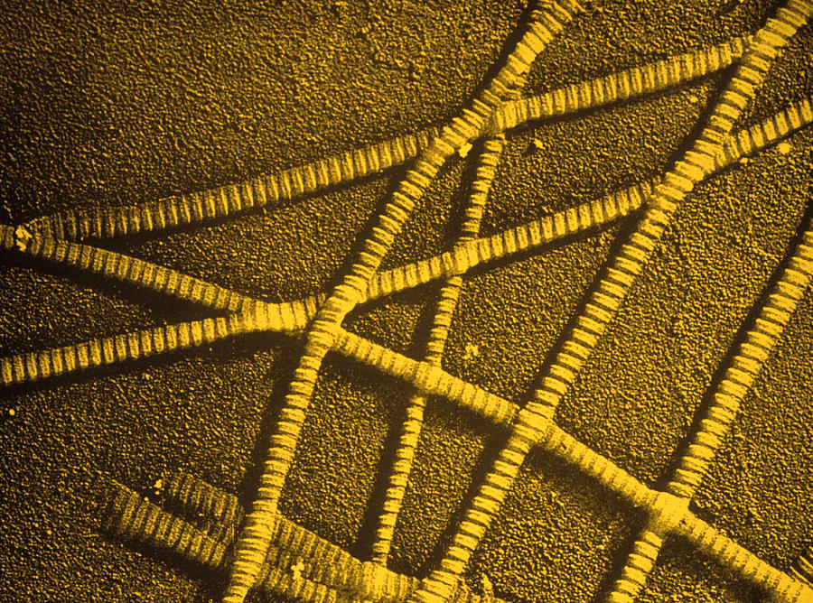
False-colour Tem Of Collagen Fibres Photograph by J Gross, Biozentrum |  Fine Art America
False-colour Tem Of Collagen Fibres Photograph by J Gross, Biozentrum | Fine Art America

TEM results of collagen mineralization after almost 1 week of reaction....  | Download Scientific Diagram
TEM results of collagen mineralization after almost 1 week of reaction.... | Download Scientific Diagram

Optical Microscopy and Electron Microscopy for the Morphological Evaluation  of Tendons: A Mini Review - Xu - 2020 - Orthopaedic Surgery - Wiley Online  Library
Optical Microscopy and Electron Microscopy for the Morphological Evaluation of Tendons: A Mini Review - Xu - 2020 - Orthopaedic Surgery - Wiley Online Library

Figure 1 from Elemental distribution analysis of type I collagen fibrils in  tilapia fish scale with energy-filtered transmission electron microscope. |  Semantic Scholar
Figure 1 from Elemental distribution analysis of type I collagen fibrils in tilapia fish scale with energy-filtered transmission electron microscope. | Semantic Scholar

File:Fibers of Collagen Type I - TEM .jpg - Wikimedia Commons

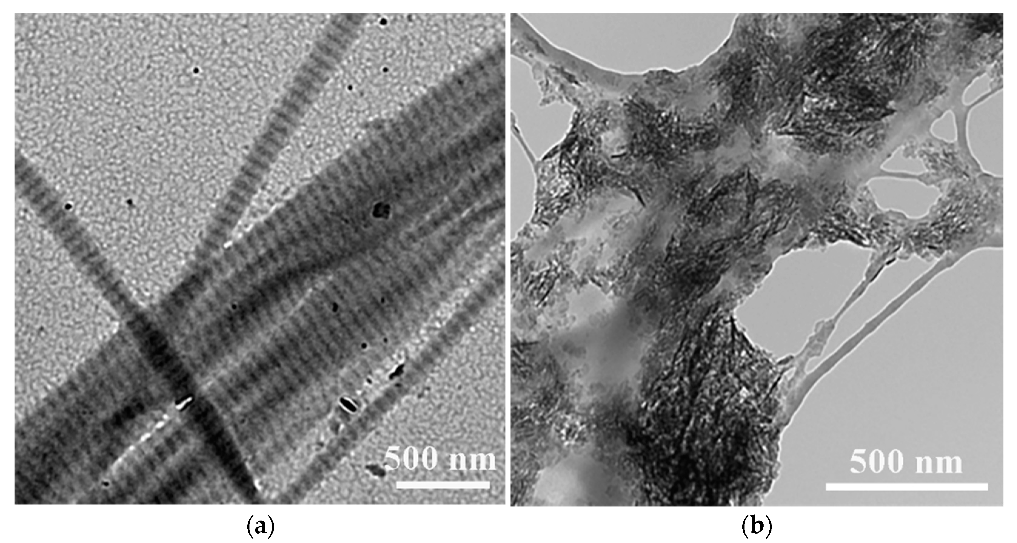
IJMS | Free Full-Text | Biomineralization of Collagen-Based Materials for  Hard Tissue Repair | HTML
IJMS | Free Full-Text | Biomineralization of Collagen-Based Materials for Hard Tissue Repair | HTML

Non-Enzymatic Decomposition of Collagen Fibers by a Biglycan Antibody and a  Plausible Mechanism for Rheumatoid Arthritis | PLOS ONE
Non-Enzymatic Decomposition of Collagen Fibers by a Biglycan Antibody and a Plausible Mechanism for Rheumatoid Arthritis | PLOS ONE

Revealing the assembly of filamentous proteins with scanning transmission  electron microscopy | PLOS ONE
Revealing the assembly of filamentous proteins with scanning transmission electron microscopy | PLOS ONE

The nanometre-scale physiology of bone: steric modelling and scanning transmission  electron microscopy of collagen–mineral structure | Journal of The Royal  Society Interface
The nanometre-scale physiology of bone: steric modelling and scanning transmission electron microscopy of collagen–mineral structure | Journal of The Royal Society Interface

Type III Collagen Can Be Present on Banded Collagen Fibrils Regardless of  Fibril Diameter
Type III Collagen Can Be Present on Banded Collagen Fibrils Regardless of Fibril Diameter

Cryo-TEM images of a collagen fiber during biomineralization. (a) At a... |  Download Scientific Diagram
Cryo-TEM images of a collagen fiber during biomineralization. (a) At a... | Download Scientific Diagram

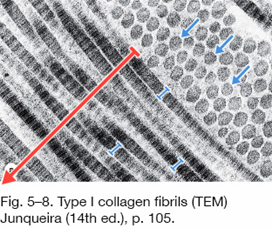
Connective Tissue Quiz Answer - Slide #7

Cryo-TEM Analysis of Collagen Fibrillar Structure - ScienceDirect